Home › Without Label › Mesothelium Histology Slide : Peritoneal Mesothelioma Unmasked By An Acute Appendicitis Gastroenterologia Y Hepatologia English Edition : Coronary vessels, nerves and adipocytes are located in this layer.
Mesothelium Histology Slide : Peritoneal Mesothelioma Unmasked By An Acute Appendicitis Gastroenterologia Y Hepatologia English Edition : Coronary vessels, nerves and adipocytes are located in this layer.
Mesothelium Histology Slide : Peritoneal Mesothelioma Unmasked By An Acute Appendicitis Gastroenterologia Y Hepatologia English Edition : Coronary vessels, nerves and adipocytes are located in this layer.. See the lining of bowman's capsule in the nephron of the kidney. The trichrome stained specimen nicely demonstrates blood vessels and dense ct in the myocardium. Function the mesothelium is composed of an extensive monolayer of specialized cells (mesothelial cells) that line the body's serous cavities and internal organs. This example is a whole mount of the mesentery stained with silver nitrate. In the skin histology slide, you will find two main layers â epidermis and dermis.
The connective tissue and the epithelium are separated by a basement membrane. slide 44 is another of example of stratified squamous keratinizing epithelium. The epicardium consists of a single layer of flattened epithelial cells, the mesothelium (m), supported by a thin layer of fibroelastic tissue (f) and adipose tissue (a). Difiore's atlas of histology with functional correlations, 12th edition.lippincott williams & Although one may divide microscopic anatomy into organology, the study of organs, histology, the study of tissues, and.
Mesothelioma Histology A Study Of Mesothelioma Cells from www.asbestos.com Pseudostratified columnar digitalscope specimen no. Its function is to support the suspension of various visceral organs within the peritoneal cavity. Your pathologist will use histology techniques to provide the most accurate information. Routine histology procedures including immunohistochemistry (ihc) and in situ hybridization (ish), protocols which can be found at our support page. Digital images for cbu histology, biol 414: 51 tongue with taste buds, monkey, he.svs 25928 x 23148 @ 40x: Move mouse coursor upon an image and you can see the image without labells. Lab activity 1 microscopic examination of epithelia 1 examine a prepared microscope slide of a whole mount of mesothelium.
Silver nitrate impregnation and haematoxylin staining, x400.
This 10 slide powerpoint presentation covers the first part of my unit on tissues for a high. This is the only slide in which the epithelium is seen from a top view. Move mouse coursor upon an image and you can see the image without labells. Pseudostratified columnar digitalscope specimen no. Note the difference in the thickness of the stratum corneum, the outermost layer. The connective tissue portion of this layer lies adjacent to the myocardium and is covered by a simple. Histopathology is the study of diseased cells. Name the layer of the heart in which purkinje fibers are found and describe their function 5. (each student should have a copy) atlas: 53 human esophagus.svs 48976 x 40839 @ 40x: The drawings of histology images were originally designed to complement the histology component of the first year medical course run prior to 2004. The stomach is a key part of the gastrointestinal (gi) tract, sitting between the esophagus and duodenum.its functions are to mix food with stomach acid and break food down into smaller particles using chemical and mechanical digestion. Notice how corrugated the cell membranes are.
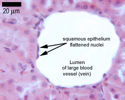
Epithelia The Histology Guide from www.histology.leeds.ac.uk The connective tissue portion of this layer lies adjacent to the myocardium and is covered by a simple. The mesothelium is a simple squamous epithelium that lines several body cavities (pleura around the lungs, pericardium around the heart, peritoneum in the abdominal cavity, and tunica vaginalis that covers the testes). Endothelium and mesothelium form cellular membranes which have the same histologic structure as Routine histology procedures including immunohistochemistry (ihc) and in situ hybridization (ish), protocols which can be found at our support page. Note that the cells that make up the epithelium have varied shapes, but that at the free surface of the epithelium they are flattened in the plane of the surface and appear elongated. Effect of the lining cells. This is the only slide in which the epithelium is seen from a top view. In a slide showing spongy bone formed solely by intramembranous ossification, all of the.
Organ / tissue slide box slide identification;
Junqueira's basic histology, 12 th ed. An introduction to cytopathology is in the cytopathology article. Silver stains the intercellular cement dark between adjacent cells. The connective tissue portion of this layer lies adjacent to the myocardium and is covered by a simple. 14 minutes the liver is the largest internal organ of the human body, weighing approximately 1.5 kg. slide 43 is typical of thick skin. Examine the major features of each of the three layers of the heart on both slides and in the images to the right. Examine this trichrome stained specimen and this h&e stained specimen of the heart wall. mesothelium = the simple squamous epithelium lining. Notice how corrugated the cell membranes are. Golgi complex in the neurons of the dorsal root ganglion. Epithelia are avascular, but all epithelia "grow" Note the difference in the thickness of the stratum corneum, the outermost layer.
Organ / tissue slide box slide identification; Histopathology is the study of diseased cells. In a slide showing spongy bone formed solely by intramembranous ossification, all of the. Endothelium and mesothelium form cellular membranes which have the same histologic structure as H 140 ward's m3304 &
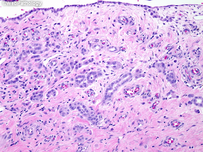
Webpathology Com A Collection Of Surgical Pathology Images from www.webpathology.com The stomach is a key part of the gastrointestinal (gi) tract, sitting between the esophagus and duodenum.its functions are to mix food with stomach acid and break food down into smaller particles using chemical and mechanical digestion. The connective tissue and the epithelium are separated by a basement membrane. 52 tooth in situ.svs 29769 x 64057 @ 40x: Use figure 6.3 to help you locate the major structures. Note the difference in the thickness of the stratum corneum, the outermost layer. Biol 414 animal histology lecture and laboratory course supplement, 2019 edition. While studying the images in this presentation, keep the. See the lining of bowman's capsule in the nephron of the kidney.
mesothelium = the simple squamous epithelium lining.
Obtain a slide of an areolar connectuve. histology is the microscopic counterpart to gross anatomy, which looks at larger structures visible without a microscope. General morphology of the cell. Note that the cells that make up the epithelium have varied shapes, but that at the free surface of the epithelium they are flattened in the plane of the surface and appear elongated. Wilkins 9781451113419 amazon.com [also acceptable: These labelled diagrams should closely follow the current science courses in histology, anatomy and Mesenteries , the sheets of connective tissue which bind together the loops of the gi tract, have the same composition as serosa and, like the serosa , and are covered on exposed faces by. Interpretation of histological sections epithelia: histology slides summary when id the tissue type, always look at the apical domain slides tissue type staining features epithelium kidney renal cortex capsule. , with differences in the layers from region to region. mesothelium = the simple squamous epithelium lining. Name the layer of the heart in which purkinje fibers are found and describe their function 5. Digital images for cbu histology, biol 414:
The drawings of histology images were originally designed to complement the histology component of the first year medical course run prior to 2004 mesothelium. June 17, 2021 reading time:


Post a Comment
Post a Comment